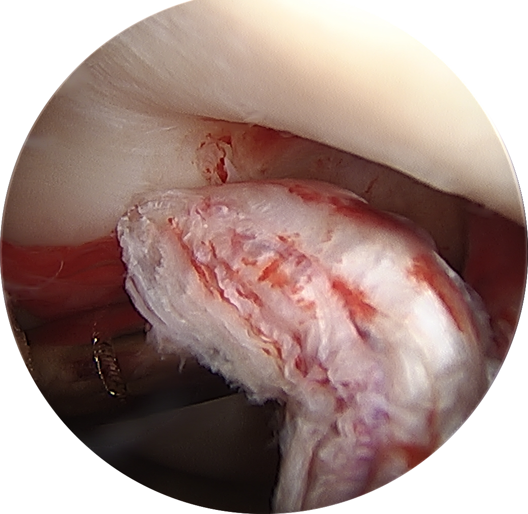
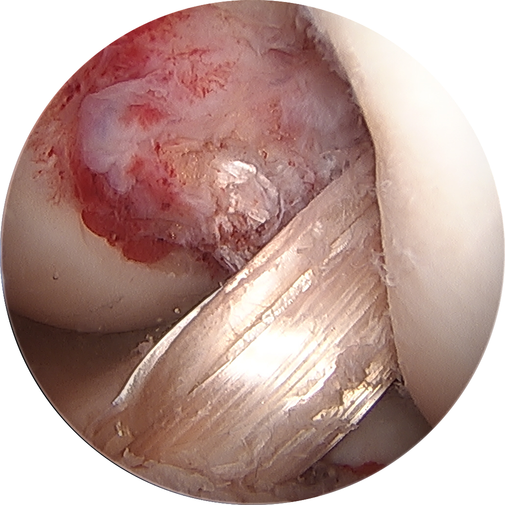
VIDEO: Vascular Trauma in Knee Injuries

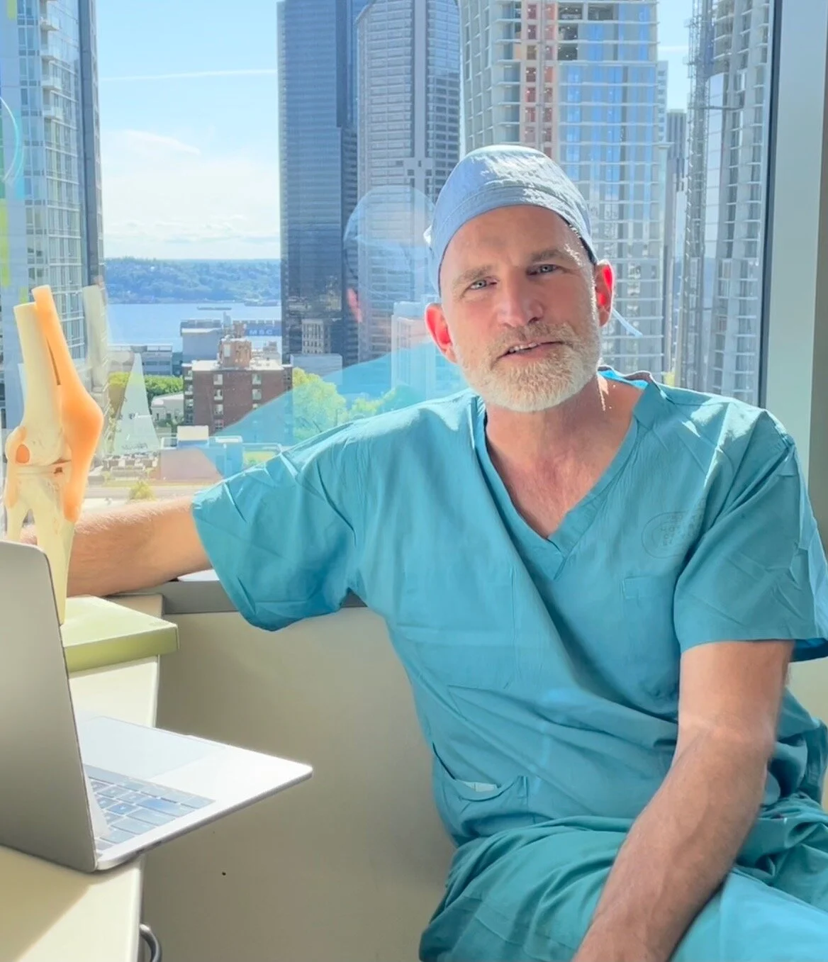
Grand Rounds at University of Washington. Discussing the importance of recognizing vascular injuries in the setting of knee trauma and the devastating limb-threatening consequences of missed vascular injuries.

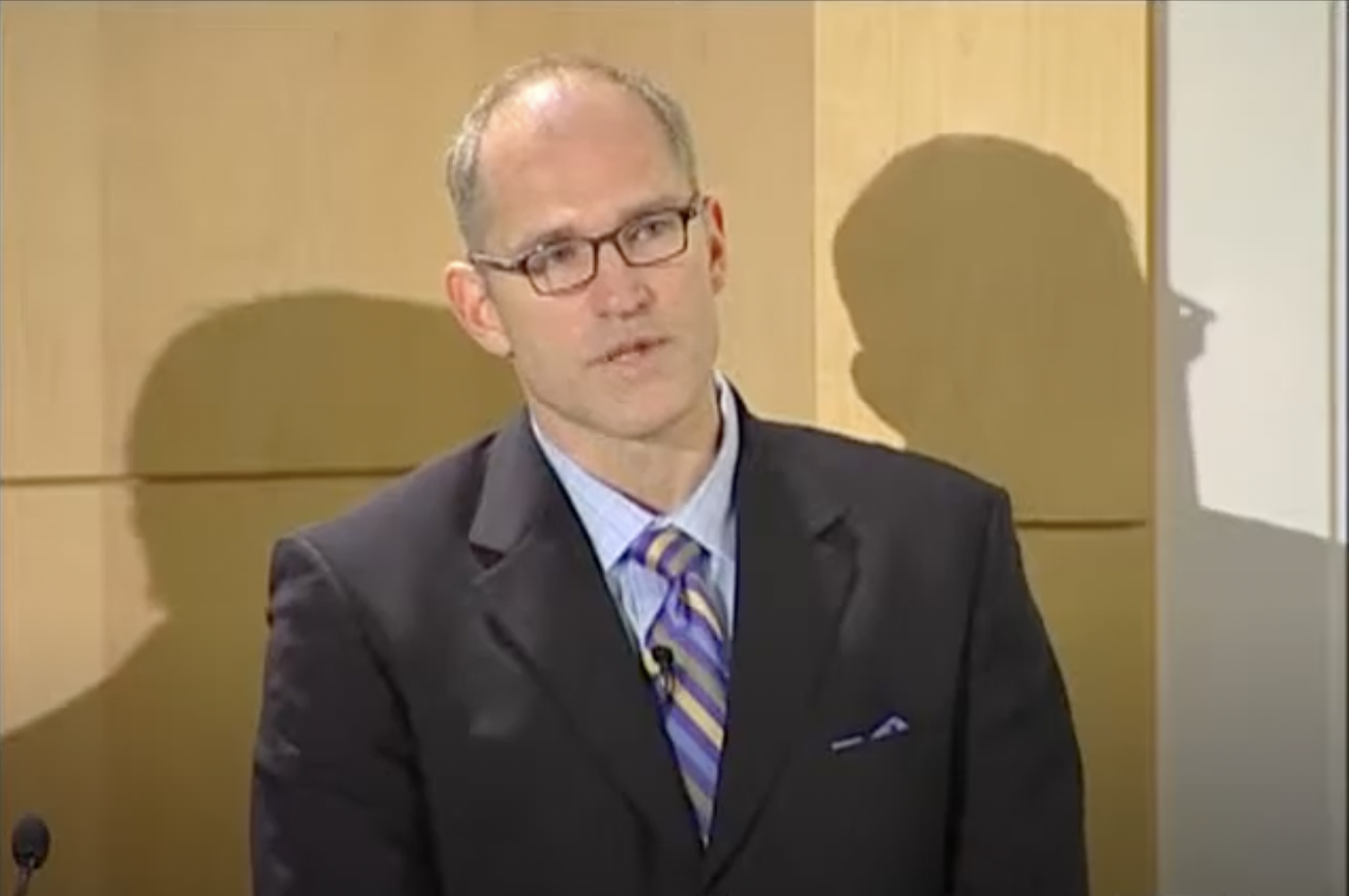
Dr. Nels E. Sampatacos, surgery resident with UW Medicine, begins the lecture with a case study along with functional anatomy concepts.

Dr. Benjamin W. Starnes, chief of vascular surgery for UW Medicine, shares his personal military experience as a vascular surgeon, providing unique insight into front-line management protocols.

Dr. Christopher J. Wahl, assistant professor with the University of Washington, discusses orthopaedic perils and pitfalls, focusing on the radiographic findings suggestive of vascular injury in the setting of knee dislocation.

Osteoarthritis = Cartilage Damage

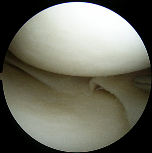
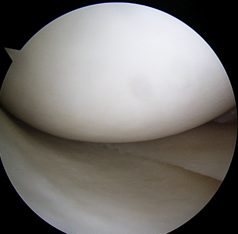
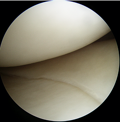
Osteoarthritis (arthritis) is the global degradation, damage or wear of articular cartilage. Articular cartilage is a smooth white tissue that covers the ends of the bones at joints. This load-bearing surface allows joints to move without friction. Unfortunately, articular cartilage has a limited capacity to heal or repair once damaged. Injury, aging, genetics or excessive loads (high repetition impact activity, obesity, etc.) can lead to cartilage degeneration. As the cartilage wears, it becomes frayed, cracked, rough and thin, thereby decreasing the protective layer of cartilage between the bones. The preservation and health of articular cartilage are paramount to joint health and the prevention of arthritis.

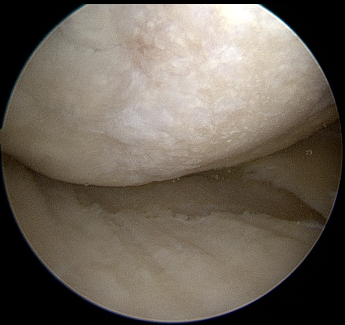
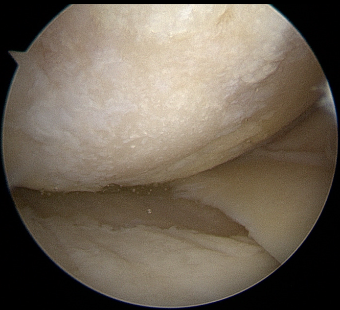
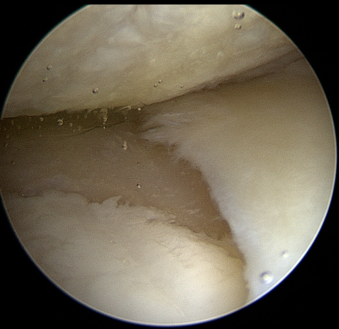
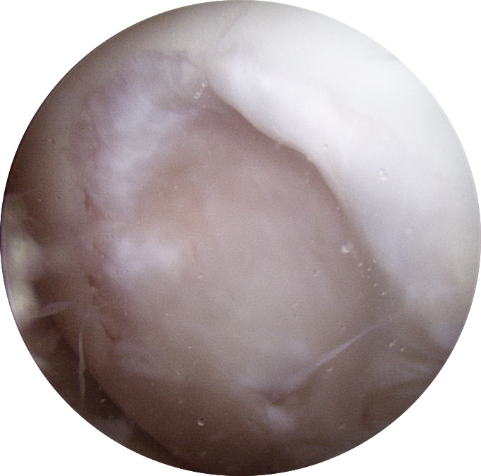
Normal Cartilage = The cartilage looks like a smooth, gliding surface; similar to a cue ball or ice rink that a zomboni just when over.


Osteoarthritis = The cartilage looks like a "gravel road"

OATs | OCA: Osteoarticular Transplant | Osteochondral Allograft

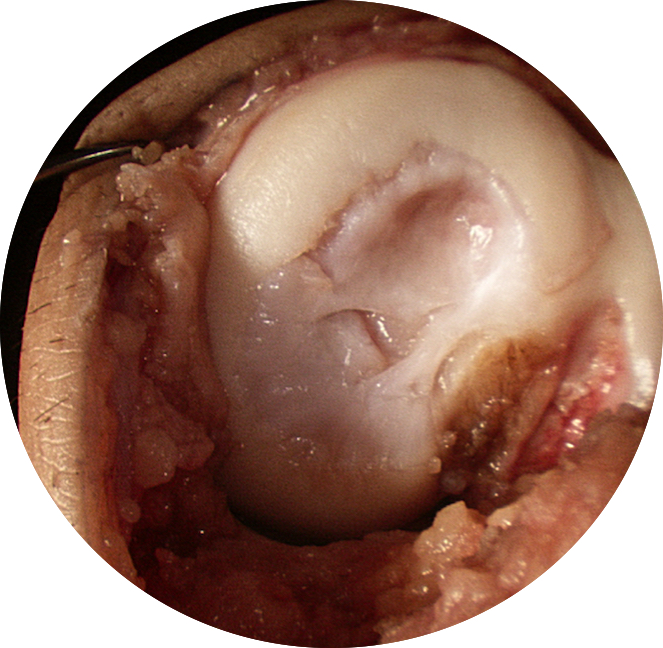
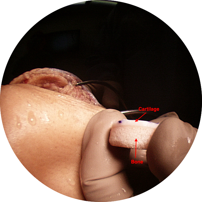
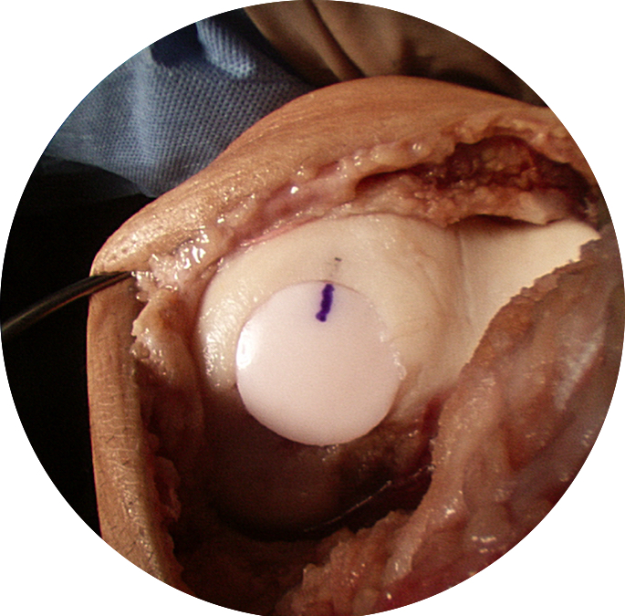
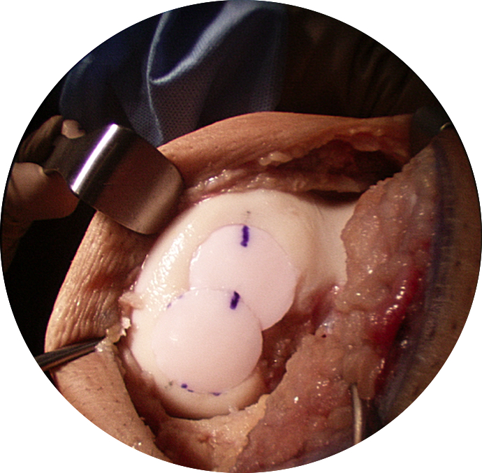
When a patient has a large cartilage defect in the knee, our preferential treatment is an OATs (osteoarticular transplant) with allograft (donor bone).  This technique allows us to resurface a large defect and match the contour of the recipient site. We prefer this procedure over others (microfracture, ACI/Carticel, DeNovo, etc) because it restores the exact architecture of the native bone-cartilage interface (subchondral bone capped with mature, hyaline cartilage with viable chondrocytes). This defect required 2 plugs, which is often called a “snowman” technique.

1️⃣Open findings of the OCD (osteochondral defect).
2️⃣Arthroscopic findings of the OCD (osteochondral defect).
3️⃣Harvested donor "plug”; showing the bone-cartilage interface. 
4️⃣The plug is advanced until flush with the surrounding native cartilage.
5️⃣A second 20 mm donor plug is placed to fill the defect.
6️⃣“Profile” of the plugs matching the contour of the native femoral condyle.

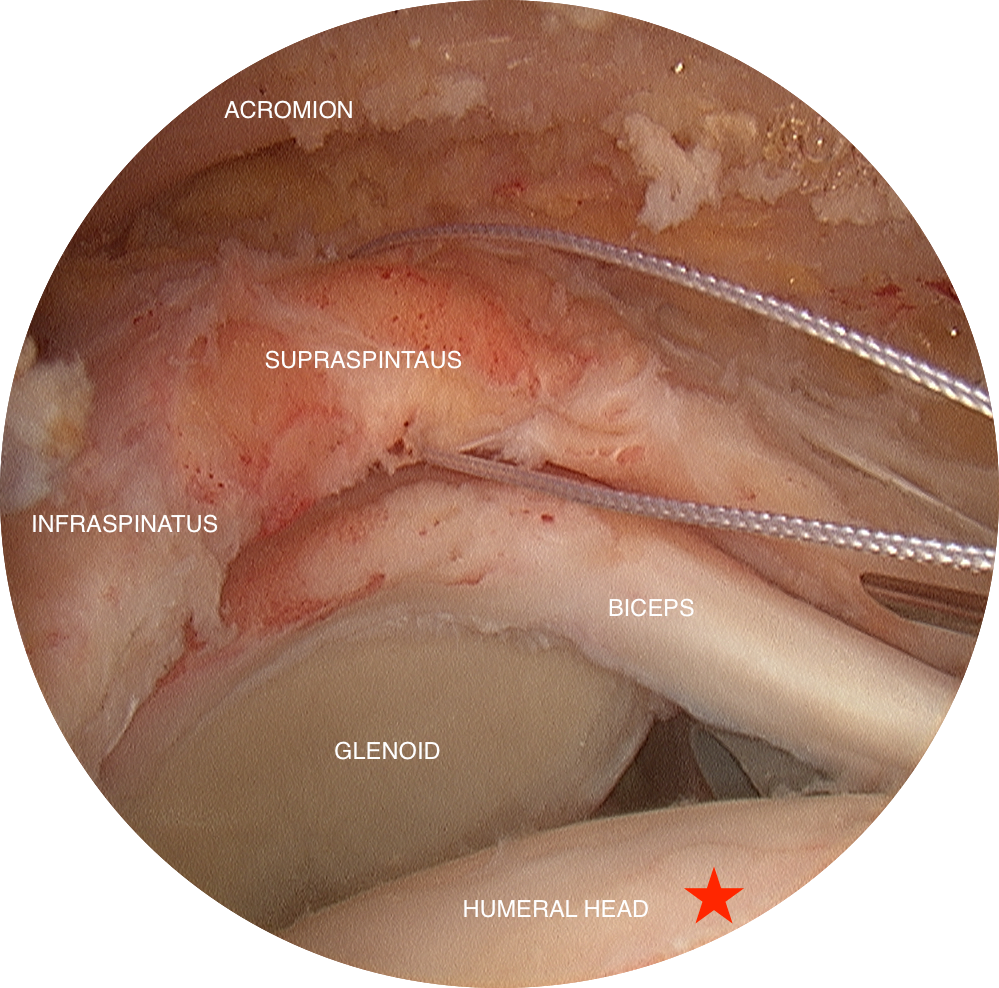
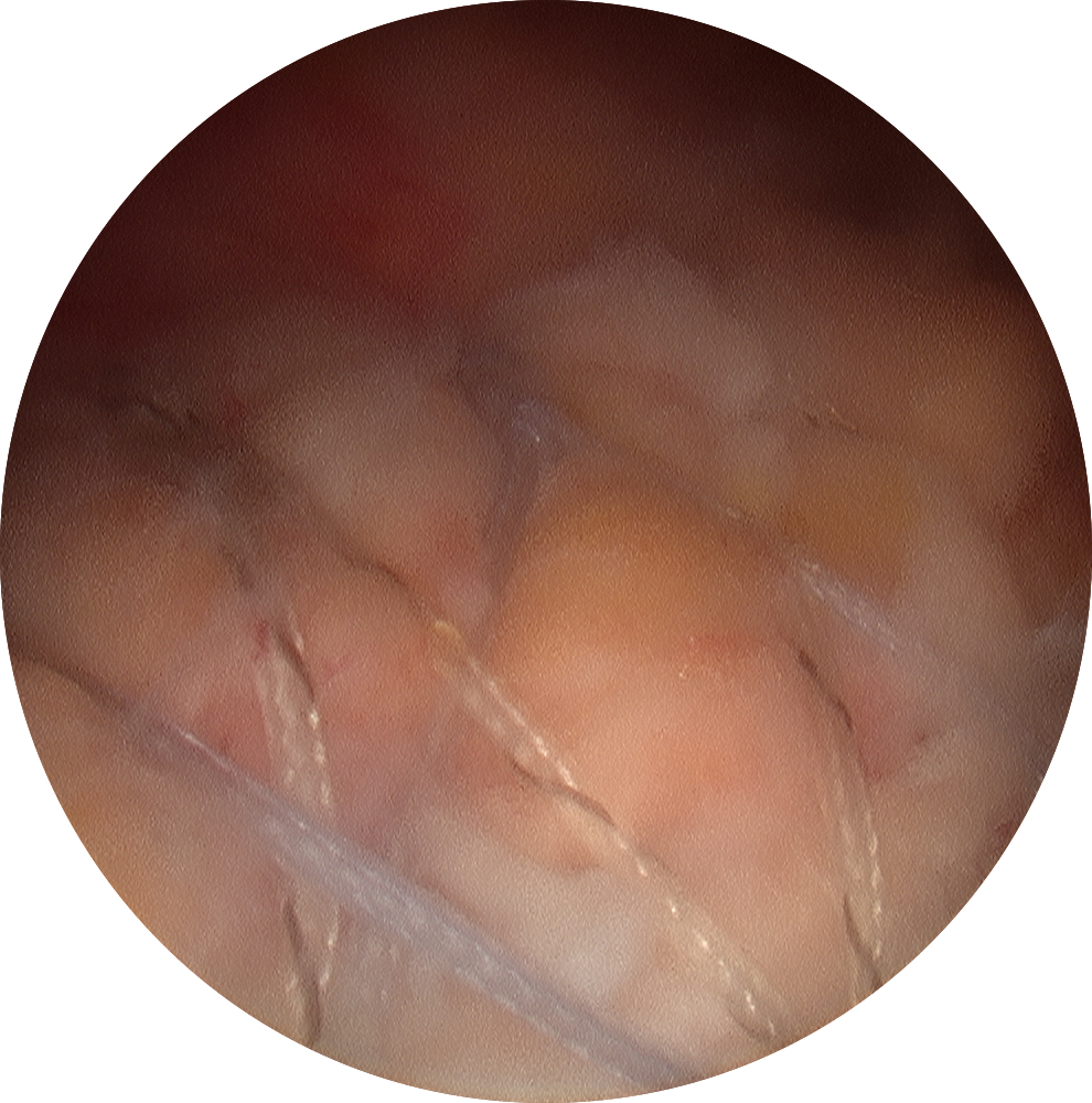
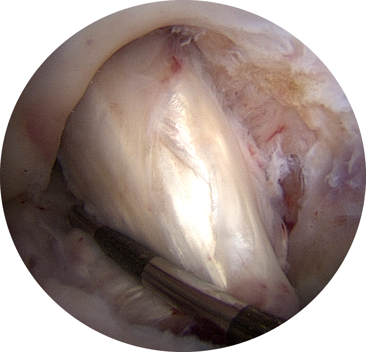
Arthroscopic Massive Rotator Cuff Repair

ch1_image_019.png

Before

This is considered a “massive” rotator cuff tear, where the supraspinatus and infraspinatus tendons have retracted back to the equator of the glenoid. In this photo, the supraspintus tendon should be attached on the greater tuberosity of the humeral head (well-beyond the red star).

ch1_image_027.png

During

Arthroscopic fixation required 3 medial (inner) row anchors (12 sutures total) and 2 lateral (outer) row anchors. The anchors are buried in the bone and then the sutures from the anchors are passed through the rotator cuff tendon. When pulling on the sutures, the cuff tendon advances to its native attachment site at the greater tuberosity.


ch1_image_029.jpg

After

As compared to the first photo, the rotator cuff tendon is now covering the great tuberosity and shoulder joint. As we say “the shoulder does not take a joke very well” and this patient will have a long recovery ahead.  Patients typically have deep, achy pain for several weeks after surgery and sleep disturbance is the #1 complaint as most prefer sleeping in a semi-reclined position or even in a reclining chair!

Extra-Articular Augmentation | Anterolateral Ligament Reconstruction

When a patient tears their native ACL and then unfortunately tears their reconstructed ACL then it is prudent to consider an ALL reconstruction (anterolateral ligament or extra-articular augmentation) when revising the intra-articular ACL.  

This structure helps control rotational instability.  We often use this as an “insurance policy”…particularly if the patient tore their graft despite having the appropriate graft and good tunnel placement.  

We also strongly consider an ALL when the patients “well” knee behaves ACL deficient with a pivot, despite having an intact ACL.

The technique is as follows…
1️⃣ Split ITB (iliotibial band).
2️⃣ Fix graft at Gerdy’s tubercle.
3️⃣ Route the graft beneath the LCL to the lateral epicondyle.
4️⃣ Find the isometric position.
5️⃣ Fix graft at isometric position.
6️⃣ Close ITB.

Extension Block from ACL Tear

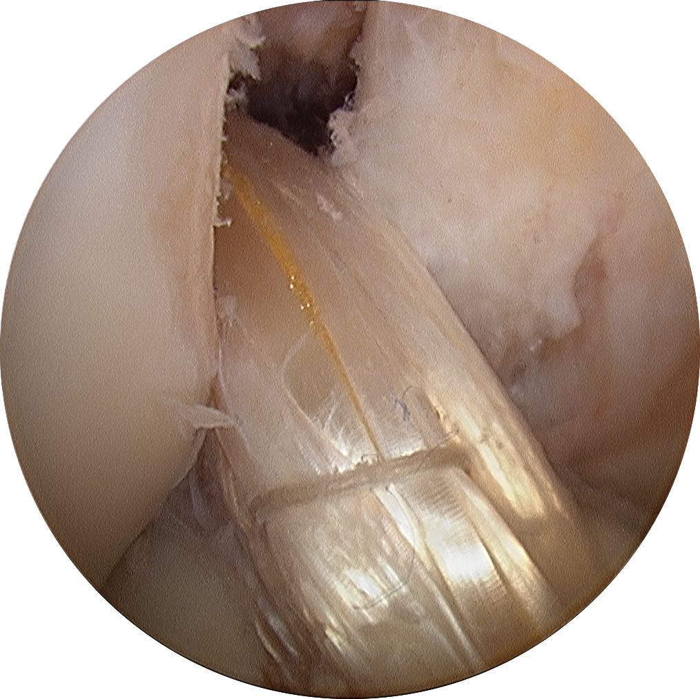
Patients with acute ACL tears can sometimes present with missing terminal extension (inability to fully straighten the knee). One must always suspect a bucket-handle meniscus tear in this situation, but sometimes it is due to “stump entrapment” where a portion of the torn ACL flips forward and gets pinched between the femur and tibia, acting as a mechanical obstruction that limits full extension of the knee.

1️⃣ + 2️⃣ Arthroscopy views of the ACL fragment caught in the intercondylar notch.
3️⃣ + 4️⃣ Sagittal and coronal MRI views of the ACL fragment flipped forward.

ACL Graft At 0 + 16 Weeks

The graft looks good....but is it ready for the patient to return to soccer?

Graft healing is an extremely complex process influenced by many variables (type of graft used, method of graft fixation + tensioning, graft motion, etc). Shockingly, during the healing process, the ACL graft tissue gradually weakens with decreased structural properties and the graft is most vulnerable at 6-7 weeks after surgery and the graft never returns to its original strength at the time of implantation!

This is why, it's important to follow your surgeons PT protocol, they know when the graft can endure more stress. No matter how good you feel or how ready you think you might be, your surgeon has a specific timeline that allows for return to running, jumping, athletics, etc. The recovery timeline is based on the biology of the graft...not how it looks on MRI, how it feels on exam or how ready you feel! Be patient...it's in your knees best interest!

Bone-Patellar Tendon-Bone (BTB or BPTB) Harvest for ACL Reconstruction

This ACL graft involves a bone plug from the tibial tubercle, the central 1/3 of the patellar tendon and a bone plug form the patella.

1️⃣ Patellar Tendon.
2️⃣ Bone plug from tibial tubercle.
3️⃣ We take a small piece of extra bone from the tibial tubercle plug and use it as bone graft in the recipient patellar bone plug site.
4️⃣ Middle 1/3 of the patellar tendon removed.
5️⃣ Closure of the patellar tendon with the knee at 90 degrees of flexion.
6️⃣ Closure of the peritenon with the knee at 90 degrees of flexion.
7️⃣ BTB autograft.
8️⃣ Final ACL reconstruction with BTB autograft.

Before + After: ACL Tear + ACL Reconstruction with Hamstring Autograft

When performing an ACL reconstruction with hamstring autograft (using the patients own hamstring tendons), 2 hamstring tendons are used to make up the graft; the semitendinosus and gracilis.  The 2 strands of tendon are sewn together to make one bundle.  Then the bundle is folded onto itself, essentially making a 4-strand graft.  

How does patient activity level influences ACL graft choice?

In a room full of orthopaedic surgeons, you can’t throw a cat without hitting a few docs who have torn their ACL’s and never had them reconstructed.  Why?  Because a lot of orthopaedists don’t regularly participate in sports that involve rapid direction change, cutting, jumping, landing or pivoting; and if you don’t do those things, the likelihood that you’ll experience knee instability is low. 

Read More

ACL Graft: Allograft

The allograft is the “Honda scooter of grafts” – It is easy to drive, you can park it anywhere, it’s dirt cheap with respect to graft pain, but they don’t work well to haul things or race, and there might be some reliability and safety issues.  You might not want it as your only vehicle.

Read More